
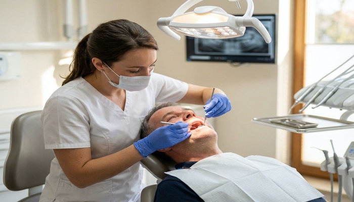
Have you ever noticed a thick, white or grayish patch inside your mouth that won’t wipe away? It could be more than just an irritation. Leukoplakia is a condition that affects the mucous membranes of the mouth and, in some cases, can signal a serious underlying health concern. While many cases are benign, a small percentage of leukoplakia patches can become cancerous — making early detection and treatment absolutely critical. Understanding what leukoplakia is, what causes it, and how it’s treated can help you take control of your oral health before a small problem becomes a bigger one.
Schedule your expert consultation at Grove Dental Clinic in Falls Church, VA today! Call (703) 578-0000 to book your appointment now!
What Is Leukoplakia?
Leukoplakia is characterized by thick, white or grayish patches that form on the tongue, gums, inside of the cheeks, or the bottom of the mouth. Unlike oral thrush (a fungal infection that also causes white patches), leukoplakia patches cannot be scraped off. The condition is most common in older adults and is often associated with tobacco use and other irritants.
There are two primary types of leukoplakia:
- Homogeneous Leukoplakia: Uniformly white patches with a flat, thin texture. This type carries a lower risk of becoming cancerous.
- Non-Homogeneous (Erythroleukoplakia): Irregular patches that may be white mixed with red areas. This type carries a significantly higher risk of malignant transformation.
A rarer but more aggressive form, called proliferative verrucous leukoplakia (PVL), spreads across a larger area and is more likely to become cancerous over time.
Common Causes of Leukoplakia
The exact cause of leukoplakia isn’t always clear, but several well-known triggers and risk factors have been identified by dental and medical professionals.
1. Tobacco Use
Smoking cigarettes, cigars, or pipes — as well as using smokeless tobacco — is the number one risk factor for leukoplakia. Chronic exposure to tobacco irritates the delicate mucous membranes inside your mouth, leading to abnormal cell growth and the formation of white patches.
2. Heavy Alcohol Consumption
Regular and excessive alcohol use can irritate oral tissues. When combined with tobacco use, the risk of developing leukoplakia — and oral cancer — increases dramatically.
3. Chronic Irritation
Rough or broken teeth, ill-fitting dentures, or damaged dental restorations can repeatedly rub against oral tissues, causing irritation over time. This is why maintaining proper dental care, including regular checkups and addressing damaged teeth quickly, is so important.
4. Human Papillomavirus (HPV)
Certain strains of HPV have been linked to the development of leukoplakia, particularly oral leukoplakia. HPV-related oral lesions are a growing concern among younger adults who do not smoke or drink excessively.
5. Weakened Immune System
People with compromised immune systems — such as those living with HIV/AIDS — are at increased risk for a related condition called hairy leukoplakia, which appears as fuzzy white patches, usually on the tongue. This form is caused by the Epstein-Barr virus.
6. Poor Oral Hygiene
Neglecting regular brushing, flossing, and professional dental cleanings can create an environment where oral tissues are more susceptible to irritation and abnormal changes. Good oral hygiene is your first line of defense against many oral health conditions, including leukoplakia.
Recognizing the Symptoms of Leukoplakia
Leukoplakia often develops gradually and may not cause any pain in its early stages. Here’s what to look for:
- White or grayish patches inside the mouth that cannot be wiped away
- Patches that may be flat and thin or thick and slightly raised
- Areas that feel hardened or tough to the touch
- Red patches mixed with white areas (a warning sign of higher cancer risk)
- Numbness, pain, or burning in the affected area (in more advanced cases)
- Difficulty chewing, speaking, or swallowing (in severe cases)
If you notice any persistent white or unusual patches in your mouth that don’t resolve within two weeks, it’s important to see your dentist or doctor as soon as possible.
Diagnosing Leukoplakia
Your dentist will typically examine the affected area during a routine dental visit. If leukoplakia is suspected, they may recommend one or more of the following diagnostic approaches:
Oral Brush Biopsy
A small brush is used to collect cells from the surface of the lesion. These cells are then analyzed under a microscope to check for abnormal or cancerous cells.
Excisional Biopsy
A small piece of tissue is surgically removed and sent to a laboratory for analysis. This is the most definitive way to diagnose leukoplakia and assess cancer risk.
Follow-Up Monitoring
In cases with low cancer risk, your dentist may recommend watchful waiting with regular monitoring appointments every three to six months.
Treatment Options for Leukoplakia
Treatment depends on the size, location, and nature of the patches, as well as whether any precancerous or cancerous cells have been detected.
Eliminating Risk Factors
The first and most important step is to stop using tobacco products and reduce or eliminate alcohol consumption. In many mild cases, removing these irritants is enough to cause the patches to disappear on their own over several weeks or months.
Dental Corrections
If leukoplakia is caused by rough teeth, broken fillings, or poorly fitting dentures, correcting these dental issues can alleviate irritation and help the patches resolve. Dental bonding may also be used to smooth out rough tooth surfaces that are causing chronic irritation.
Medications
In some cases, doctors may recommend topical treatments or systemic medications, including antiviral drugs (for hairy leukoplakia associated with HIV) or retinoid therapy in select patients. However, medication effectiveness varies, and recurrence is common.
Surgical Removal
If a biopsy reveals precancerous or cancerous cells, surgical removal of the patches is typically recommended. This can be done through:
- Conventional Surgery: Cutting away the lesion using a scalpel.
- Laser Therapy: Using a focused laser beam to precisely remove the affected tissue with minimal bleeding and scarring.
- Cryotherapy: Freezing the abnormal tissue to destroy it.
- Photodynamic Therapy: Using light-activated drugs to destroy abnormal cells.
Ongoing Monitoring
Even after successful treatment, leukoplakia can recur, especially if tobacco or alcohol use continues. Regular dental checkups — at least twice a year — are essential for early detection of any recurrence.
Preventing Leukoplakia: What You Can Do
Prevention is always better than treatment. Here are proactive steps you can take to significantly reduce your risk of developing leukoplakia:
- Quit tobacco: This is the single most effective way to reduce your risk.
- Limit alcohol: Moderate your intake and avoid combining alcohol with tobacco.
- Practice excellent oral hygiene: Brush twice daily, floss every day, and rinse with an antibacterial mouthwash.
- Eat a healthy diet: A diet rich in fruits and vegetables provides antioxidants that help protect oral tissues.
- Schedule regular dental visits: Professional exams allow early detection of any suspicious changes in your mouth.
- Address dental issues promptly: Fix cracked teeth, rough fillings, or ill-fitting dentures quickly to prevent chronic irritation.
When to See a Dentist
Don’t wait until you’re in pain to see your dentist. Any white patch, sore, or unusual change in your mouth that persists for more than two weeks deserves professional attention. Early intervention is the key to preventing leukoplakia from progressing to something more serious.
Your dentist is your partner in oral health. Regular appointments aren’t just about cleaning your teeth — they’re about safeguarding your overall well-being. Make sure leukoplakia screening is part of your routine dental checkup conversation.
FAQ
Is leukoplakia the same as oral cancer?
No, leukoplakia is not the same as oral cancer. However, it is considered a potentially precancerous condition. A small percentage of leukoplakia lesions — particularly the non-homogeneous type — can develop into oral cancer over time. That’s why prompt evaluation and monitoring are so important.
Can leukoplakia go away on its own?
In some mild cases, especially when caused by tobacco or irritation, leukoplakia patches may resolve on their own once the irritant is removed. However, not all cases resolve without treatment, and a dental professional should always evaluate persistent patches.
Is leukoplakia contagious?
Standard leukoplakia is not contagious. However, hairy leukoplakia — associated with the Epstein-Barr virus in individuals with HIV — involves a virus that can be transmitted, though the condition itself is not spread through casual contact.
How long does it take for leukoplakia to develop?
Leukoplakia typically develops gradually over time, often as a result of years of tobacco use or chronic irritation. It can take months or even years before noticeable patches form.
Can children get leukoplakia?
Leukoplakia is rare in children and is far more common in adults over 40. Cases in younger individuals may be linked to HPV or other specific causes and should be evaluated promptly.
Does flossing help prevent leukoplakia?
Flossing is an essential part of good oral hygiene and helps reduce bacterial buildup and gum irritation. While flossing alone won’t prevent leukoplakia, it contributes to an overall healthier oral environment that is less susceptible to abnormal tissue changes.
What does leukoplakia look like?
Leukoplakia typically appears as thick, white or grayish patches inside the mouth. These patches may be flat, slightly raised, or have an irregular texture. Unlike milk residue or oral thrush, they cannot be wiped away. Some patches may include red areas, which can indicate a higher risk of malignancy.
Schedule your expert consultation at Grove Dental Clinic in Falls Church, VA today! Call (703) 578-0000 to book your appointment now!


